Heart Disease
Do you have any of these symptoms?
- Chest pain or discomfort
- Tightness in the chest and jaw and/or nausea during physical exertion
- Sudden shortness of breath after physical activity
- Lightheadedness or breaking out in a cold sweat for no apparent reason
- Other warning signs may signal potentially life-threatening heart rhythm problems. Suddenly passing out without virtually any warning of any sort is an indication that there may be a serious rhythm problem.
- a family history of heart disease or unexplained sudden death
- smoking
- diabetes
- high cholesterol
- high blood pressure
- obesity or overweight
- physical inactivity
- advancing age, especially men >45 and women >55
New study for evaluation of coronary artery disease
CT angiography refers to a computed tomography (CT) exam that is specially designed to evaluate the coronary arteries. CT angiography is a relatively new technique in the evaluation of coronary arteries that has recently become available due to technical advances in computed tomography, notably 64-slice CT. 64 slice CT refers to scanners that have 64 rows of detectors instead of just one row. This allows for higher quality images because more of the heart can be scanned with each rotation of the scanner. The scans can also be performed in a shorter amount of time. Prior to 64-slice computed tomography, these studies did not produce quality images. The main problem is that unlike other parts of the body, the heart is in constant motion. The coronary arteries are very small, and this small size combined with the motion of the heart made it difficult to evaluate the coronary arteries. With new generation 64-slice computed tomography, images of the entire heart are rapidly created. 64-slice CT angiograms are less invasive than traditional catheter-based angiograms. A catheter based angiogram uses a catheter to inject dye into a large artery or vein, and the heart is then visualized by x-ray technology. Catheter angiograms provide better resolution than CT angiograms, though they are more invasive and prone to complications. CT angiograms can be used to evaluate areas of individual coronary arteries where the accumulation of plaque has led to narrowing of the artery. CT angiography can evaluate areas of narrowing due to calcified plaque, as well as soft plaques that do not have calcium and are frequently the cause of sudden heart attacks.
Heart research shifts to early detection
By Stephen Smith : Boston Globe : November 6, 2006
It is one of the most enduring and intractable problems in medicine: In half of men and two-thirds of women who die abruptly from heart disease, there is no warning, no symptom of a problem brewing.
Now, scientists from Boston to Tokyo are intensifying a high-stakes race to better understand the phenomenon and come up with a way to detect trouble deep inside the heart. They are using sound and light to peer directly into the heart's vessels, hunting for clumps of fat not much larger than a grain of rice and primed to rupture.
The experimental detection systems are all the talk at major meetings of cardiologists, with preliminary findings presented last month at a conference in Washington and more reports expected next week at a Chicago convention.
"It's like a mystery novel -- we're trying to recreate the crime looking backwards in time. You have the smoking gun, which is a heart attack," said Dr. Sergio Waxman, a cardiovascular researcher at Lahey Clinic and leader of a plaque-detection trial. "The question is, how do we prevent this from happening?"
If one of the approaches under development succeeds -- and that's not guaranteed -- the consequences could be profound, for patients and scientists alike. It could translate into tens of thousands of lives saved annually. And it could have equally staggering economic implications, with one market analyst estimating that detection systems have the potential to generate billions of dollars in revenue, dwarfing recent innovations such as stents.
But the efforts to detect dangerous deposits of fat also have reinforced long-standing rifts in one of the most competitive and lucrative realms of medicine.
One camp of cardiovascular specialists argues that heart disease can't be stopped without looking at the patient's entire coronary system. They remain deeply skeptical about the ability of any test to identify the precise location of a potential problem, and even if researchers develop such a test, the doubters ask: What would we do with that information?
"We need to draw a very careful line about raising hopes of the public that there will be some wonderful gizmo that will allow us to put the finger in the dike," said Dr. Peter Libby, chief of cardiovascular medicine at Brigham and Women's Hospital. "We could save tens of thousands of heart attacks if we applied what we already know -- like the current guidelines on cholesterol, blood pressure, diet. But these are things that are boring to the public."
Another camp -- and that's the camp working on the detection systems -- emphasizes the importance of tracking down those fat deposits believed to be most likely to rupture and create fatal blockages. Champions of that approach have a name for those deposits: vulnerable plaque.
"What I generally say is I agree it's a systemic disease, and it is very important to treat it systemically," said Dr. James E. Muller, chief executive of a Burlington biotech company called InfraReDx, which has attracted $50 million from investors to create a plaque-detection system. "But, unfortunately, if you go into a coronary care unit, you'll find it's full of people who've been taking their statins to lower their cholesterol and even taking an aspirin -- the best systemic therapy possible -- and they still have a heart attack."
And when that patient undergoes surgery to open a blockage, Muller said, "you find there's one spot in the artery that caused the clot."
To locate that spot, researchers must travel directly into the heart because sufficiently detailed snapshots can't be taken through skin, bone, and muscle.
There's nothing new about shooting images inside the heart, of course: Specialists do that routinely while clearing blocked vessels during a procedure called angioplasty. But while the picture-taking systems used during those operations are good at spying big blockages, they're not so good when it comes to examining the contents of smaller, rupture-prone plaques, said Dr. Momtaz Wassef of the National Heart, Lung, and Blood Institute.
So researchers have enlisted some tried-and-true technologies, including ultrasound and infrared, to attempt to identify troublesome fat deposits in what Muller described as a "harsh environment" for such testing: the beating human heart. They're not looking for major blockages. Instead, they're hunting for plaques with the right mix of fat and a thin cap on top that make them vulnerable to rupture.
Thousands of patients around the world are participating in studies of these detection systems.
A California company called Volcano, for example, has enrolled 3,000 patients in a trial of its advanced ultrasound system, said Vince Burgess, the company's marketing vice president.
Another technique measuring echoes of light -- optical coherence tomography -- has been tried on about 150 patients at Massachusetts General Hospital by Dr. Ik-Kyung Jang. A Westford company called LightLab is selling the optical systems in Europe, China, and South Korea, and US trials are almost finished, said Joe Schmitt, the firm's chief technology officer.
Muller's InfraReDx has also begun a trial of its system, which uses infrared laser light to read the chemical fingerprints of plaques. The trial ultimately will include about 100 patients at five hospitals, including Lahey in Burlington.
InfraReDx has received preliminary FDA approval to test its method, but that only means it's acceptable to use the technique in human arteries; it says nothing about the system's ability to detect fatty deposits. Muller said the company intends to seek approval for that more-specific use in March, and hopes to begin selling units next summer.
"I take it for granted that one or more of these techniques will work," said Dr. Gary Mintz, director of publications of the Cardiovascular Research Foundation, a private medical research organization. "But they do have some risks. There are complications associated with every technique. So, the question is, is it worth it?"
Use of plaque-detection systems would be limited to patients already undergoing invasive procedures, such as angioplasty. Such patients, who have had a life-threatening heart problem, are at dramatically higher risk of another one, and, developers of the systems argue, could especially benefit from detection of dangerous fat deposits.
The market could be substantial: Muller said 1 million US patients and 2 million worldwide annually undergo procedures to clear clogged arteries.
Jan Wald, a medical device analyst at the financial services firm A.G. Edwards & Sons who is familiar with the competing plaque-detection systems, said that "in the investment community, the question is: How quick are the techniques going to be utilized and available? Is it going to be realized in 2007? 2012? 2020?"
But even if scientists succeed in perfecting a system to pinpoint dangerous fat deposits, there's no agreement on what to do about them. Would it be enough to give patients high doses of cholesterol-lowering drugs? Would it be necessary to implant tiny metal scaffolds to keep arteries open?
"Are you going to put three, four, five stents in people with vulnerable plaque?" Jang asked. "I would not."
At medical conferences, the elite of cardiology continue to joust over plaque-detection systems, with true believers facing off against naysayers such as Dr. Steven Nissen, chairman of cardiovascular medicine at the Cleveland Clinic.
"They're barking up the wrong tree," Nissen said. "This has been an elusive target and the chance that any one of these little companies is going to hit a home run here after 15 years of all this discussion is, I think, relatively low.
"Would I like someone to hit a home run and help us figure out where to intervene? The answer, of course, is yes."
Weighing the Costs of a CT Scan’s Look Inside the Heart
By Alex Berenson and Reed Abelson
NY Times Article : June 29, 2008
A group of cardiologists recently had a proposition for Dr. Andrew Rosenblatt, who runs a busy heart clinic in San Francisco: Would he join them in buying a CT scanner, a $1 million machine that produces detailed images of the heart?
The scanner would give Dr. Rosenblatt a new way to look inside patients’ arteries, enable his clinic to market itself as having the latest medical technology and provide extra revenue.
Although tempted, Dr. Rosenblatt was reluctant. CT scans, which are typically billed at $500 to $1,500, have never been proved in large medical studies to be better than older or cheaper tests. And they expose patients to large doses of radiation, equivalent to at least several hundred X-rays, creating a small but real cancer risk.
Dr. Rosenblatt worried that he and other doctors in his clinic would feel pressure to give scans to people who might not need them in order to pay for the equipment, which uses a series of X-rays to produce a composite picture of a beating heart.
“If you have ownership of the machine,” he later recalled, “you’re going to want to utilize the machine.” He said no to the offer.
And yet, more than 1,000 other cardiologists and hospitals have installed CT scanners like the one Dr. Rosenblatt turned down. Many are promoting heart scans to patients with radio, Internet and newspaper ads. Time magazine and Oprah Winfrey have also extolled the scans, which were given to more than 150,000 people in this country last year at a cost exceeding $100 million. Their use is expected to soar through the next decade. But there is scant evidence that the scans benefit most patients.
Increasing use of the scans, formally known as CT angiograms, is part of a much larger trend in American medicine. A faith in innovation, often driven by financial incentives, encourages American doctors and hospitals to adopt new technologies even without proof that they work better than older techniques. Patient advocacy groups and some doctors are clamoring for such evidence. But the story of the CT angiogram is a sobering reminder of the forces that overwhelm such efforts, making it very difficult to rein in a new technology long enough to determine whether its benefits are worth its costs.
Some medical experts say the American devotion to the newest, most expensive technology is an important reason that the United States spends much more on health care than other industrialized nations — more than $2.2 trillion in 2007, an estimated $7,500 a person, about twice the average in other countries — without providing better care.
No one knows exactly how much money is spent on unnecessary care. But a Rand Corporation study estimated that one-third or more of the care that patients in this country receive could be of little value. If that is so, hundreds of billions of dollars each year are being wasted on superfluous treatments.
At a time when Americans are being forced to pay a growing share of their medical bills and when access to medical care has become a major political issue for states, Congress and the presidential candidates, health care experts say it will be far harder to hold down premiums and expand insurance coverage unless money is spent more wisely.
The problem is not that newer treatments never work. It is that once they become available, they are often used indiscriminately, in the absence of studies to determine which patients they will benefit.
Some new treatments, like the cancer drug Gleevec and implantable heart defibrillators, undoubtedly save lives, contributing to the United States’ reputation for medical breakthroughs. But others — like artificial spinal disks, which can cost tens of thousands of dollars to implant but have not been shown to reduce back pain in many patients, and Vytorin, a new cholesterol drug that costs 20 times as much as older medicines but has not been proved superior — have been criticized for not justifying their costs.
And sometimes, the new technologies prove harmful. Physicians were stunned, for example, when clinical trials showed last year that expensive anemia medicines might actually hasten death in kidney and cancer patients. Such drugs are used more widely in the United States than elsewhere.
“We have too many situations where we thought we knew what the answer was and it didn’t turn out like everyone thought,” said Dr. Mark Hlatky, a cardiologist and professor of health research and policy at Stanford University.
A Tool of Dubious Value
The problem of inadequate study is especially serious for medical devices and imaging equipment like scanners, which typically are not as strictly regulated as prescription drugs. Under Food and Drug Administration regulations, the makers of CT scanners — CT is short for computed tomography — do not have to conduct studies to prove that their products benefit patients, as drug makers do. The manufacturers must certify only that the scanners are safe and provide accurate images.
Once the F.D.A. approves a test or device, Medicare rarely demands evidence that it benefits patients before agreeing to pay for it. But last year, Medicare officials raised questions about the benefits of CT heart scans and said it would demand more studies before paying for them. But after heavy lobbying by cardiologists, Medicare backed down. Private insurers, while initially reluctant to pay for the tests, are also covering them.
Physicians in this country have a free hand in deciding when to use new technology like CT angiography. Some are conservative. But others, especially doctors in private practice who own their scanners, use the tests aggressively.
Douglas Ring, a 63-year-old Los Angeles real estate developer, said he received a CT heart scan in October 2005, on the advice of Dr. Ronald P. Karlsberg, a Beverly Hills cardiologist. “Ron has been my physician for 15 or 20 years, and he got this new toy in his office, and he said I should try it,” Mr. Ring said. He took the test despite having no symptoms of heart disease, like shortness of breath and chest pain. He was already taking cholesterol medicine, and a different test had shown no problems with his heart.
The CT heart scan by Dr. Karlsberg found a moderate buildup of plaque in one of Mr. Ring’s coronary arteries. The doctor increased Mr. Ring’s cholesterol medicines and encouraged him to diet and exercise.
Dr. Karlsberg said he considered the information from Mr. Ring’s CT scan extremely valuable. “Here’s a case of near-serious coronary disease that required medical management,” said Dr. Karlsberg, a partner at the Cardiovascular Medical Group of Southern California, which conducted about 1,400 CT heart scans last year.
Many other cardiologists, though, say patients like Mr. Ring do not benefit from CT scans. And by the time they are 50, most people will have plaque visible on a CT scan, so the findings of Mr. Ring’s scan were not surprising.
Arteries narrowed by plaque are not necessarily a threat, said Dr. Eric Topol, a practicing cardiologist and director of the Scripps Translational Science Institute in La Jolla, Calif. The danger arises when bits of plaque break and produce a clot that blocks blood to the heart. But CT angiograms cannot tell whether a particular blockage is likely to rupture or, except in extreme cases, is keeping the heart from receiving enough blood.
If doctors do choose to treat blockages, they can insert stents — small metal scaffolds that prop open arteries. But while stents have been proved to reduce chest pain, they have not been shown to prolong patients’ lives or help them avoid heart attacks. Patients with the most severe blockages can receive bypass surgery, which when necessary can be a lifesaving procedure.
And so patients who do not have chest pain, like Mr. Ring, should not receive CT heart scans, said Dr. Rita Redberg, a cardiologist and researcher at the University of California, San Francisco, who is a leading critic of the scans.
“No data suggests that there’s any reason for anyone asymptomatic to have a test,” she said. “There certainly is this idea that having a test can help you prevent a heart attack, and I don’t know where it came from.”
Further, each scan creates an additional lifetime risk of cancer that is somewhere between 1 in 200 and 1 in 5,000, said Dr. David J. Brenner, director of the Center for Radiological Research at Columbia University. Younger patients and women are at higher risk.
Dr. Karlsberg and other cardiologists who support widespread use of CT heart scans argue that they can reduce the need for other tests — like conventional angiograms, which can find plaque but require a catheter to be threaded through the arteries. Conventional angiograms are more expensive than CT scans and carry their own risks.
If a CT heart scan finds plaque that a doctor intends to treat with a stent, a conventional angiogram will still be necessary to determine where and how to implant the stent. So a CT scan does not always eliminate the need for a conventional angiogram.
The most valuable use of a CT angiogram may be when a patient comes to an emergency room complaining of chest pains but has few other symptoms of a heart attack. The test can quickly rule out heart trouble. But such patients represent a minority of those receiving CT heart scans.
Dr. Karlsberg also pointed to the case of a seemingly healthy 68-year-old patient whom he scanned in his office in 2006. To the shock of both doctor and patient, the scan revealed a 95 percent blockage of the patient’s main coronary artery. The patient had immediate bypass surgery to relieve the blockage, an operation that may have saved his life, Dr. Karlsberg said. The man, who cited privacy concerns in asking that his name not be used, confirmed the doctor’s account.
Cardiologists who oppose wide use of the scans agree that they can sometimes find dangerous blockages that require immediate surgery in asymptomatic patients. But they said such cases are extremely rare — not common enough to justify using the scans routinely, given their cost and radiation risks.
For too many people, the scans are simply inappropriate, said Dr. Howard C. Herrmann, director of interventional cardiology at the University of Pennsylvania. “I find many patients have CT angiograms who shouldn’t be getting CT angiograms.”
As more than 13,000 heart doctors gathered in Chicago in late March for the annual American College of Cardiology conference, the biggest and best-located booths belonged to General Electric, Philips Electronics, Siemens and Toshiba, the leading makers of the machines used for CT angiograms.
Cardiologists hired by the companies offered short briefings on ways to reduce radiation doses, while sales representatives in business suits quietly talked up the benefits of the scans and the clarity of the images. The sales atmosphere was low key, more art gallery than “Glengarry Glen Ross.”
A hard sell is unnecessary because the manufacturers are finding a receptive audience. Many cardiologists have been eager for a new tool that lets them see inside the heart with unprecedented clarity — while also providing a new source of revenue.
Use of CT scans accelerated after 2004, when manufacturers introduced a new generation called 64-slice scanners, which are fast enough to capture images of a beating heart. The scanners fire X-rays in a series of rotations around the torso, generating thousands of narrow vertical images. Sophisticated software then combines data from the X-rays into a single image.
The Financial Incentives
Already, more than 1,000 hospitals and an estimated 100 private cardiology practices own or lease the $1 million CT scanners, which can be used for the angiograms and for other imaging procedures. Once they have made that investment, doctors and hospitals have every incentive to use the machines as often as feasible. To pay off a scanner, doctors need to conduct about 3,000 tests, industry consultants say.
Fees from imaging have become a significant part of cardiologists’ income — accounting for half or more of the $400,000 or so that cardiologists typically make in this country, said Jean M. Mitchell, an economist at Georgetown University who studies the way financial incentives influence doctors.
Besides generating profits themselves, the scans enable cardiologists to find blockages in patients who have no symptoms of heart problems. Doctors can then place stents in patients who would not otherwise have received them, generating additional revenue of $7,500 to $20,000 per patient.
While clinical trials have not shown that stents benefit patients with no symptoms of heart disease, they are still routinely inserted in such patients when tests find significant blockages. Cardiologists joke that the phenomenon is “ocular stenosis” — blockages that can be seen are stented.
“You find a lot of asymptomatic disease,” said John O. Goodman, a business consultant to cardiologists. “It will put more patients in the cath lab” — medical shorthand for a cardiac catheterization laboratory, where conventional angiograms and stenting procedures take place.
Ms. Mitchell said cardiologists simply practice medicine the way the health system rewards them to. Given the opportunity to recommend a test for which they will make money, the doctors will.
“This is not greed,” she said. “This is normal economic behavior.”
Doctors who perform a lot of CT heart scans tend to be evangelists for the technology. Dr. John A. Osborne, a cardiologist in solo practice in Grapevine, Tex., just outside Dallas, argues that CT angiograms catch heart disease at its earliest stages. His Web site, sothcardiology.com, states the proposition in stark terms: “Half of Americans have died of heart attacks and strokes. Which one are you?”
Supported by a staff of about 20 people, Dr. Osborne estimates that he does 15 CT angiograms a day. Arterial plaque is “cancer of the coronaries,” he tells patients. “Do you have it or not?”
Before their plaque creates symptoms, Dr. Osborne asserts, patients should be aggressively treated, urged to diet and exercise and given cholesterol-lowering and other drugs.
Scans ‘Sell Themselves’
Like many cardiologists who perform CT scans, Dr. Osborne relies on primary-care doctors to send him candidates. He frequently gives lectures to primary-care doctors on the technology’s benefits. When doctors see the images, he said, “they become true believers.”
Two years ago, Dr. Osborne persuaded a family practice doctor, Dr. Michael Dotti, to have his own CT angiogram at no cost. Dr. Dotti was amazed at the scan’s ability to spot early signs of disease. “It’s nice to know I have clear arteries at 51,” he said. “The scans sort of sell themselves.”
The technology has been covered in the news media, including a September 2005 Time magazine cover on CT angiograms, “How New Heart-Scanning Technology Could Save Your Life.” The following month, Oprah Winfrey devoted a segment of her television program to women’s heart disease and recommended that her viewers consider taking the test. Representatives for Time and Ms. Winfrey declined to comment on their coverage of the technology.
Even cardiologists who think the CT scans are overused say they may one day prove valuable. If manufacturers can produce scanners that can determine which plaques are stable and which are likely to rupture, the machines could revolutionize the treatment of heart disease. Patients found to be at low risk might be able to avoid taking medicine entirely, while others would be given intensive treatment.
But for now, doctors cannot use the images that way. Finding out whether the heart is actually short of blood and at high risk for an attack requires tests other than a CT scan — most likely, a nuclear stress test, which uses radioactive dye to track blood flow through the coronary arteries.
The CT angiogram is “a great technology searching for a great application,” said Dr. Charanjit S. Rihal, the director of the cardiac catheterization laboratory at the Mayo Clinic in Rochester, Minn., who sees little diagnostic value in the current generation of heart scanners.
CareCore National, a Bluffton, S.C., company that reviews treatment and test requests for health insurers, has found that when doctors request a CT angiogram for a patient, they also frequently ask for a nuclear stress test.
“We’re seeing layering of tests on top of each other,” said Dr. Russell Amico, a CareCore executive. His company denies as many as 70 percent of the CT scans requested, a much higher rate of rejection than for other kinds of tests his company reviews.
Impatient Patients
Sometimes, it is not the doctor but the patient who is eager for the scan. On a recent Wednesday morning on the Upper East Side of Manhattan, Dr. Harvey Hecht at Lenox Hill Hospital watched from a lead-shielded control room as a 59-year-old patient, Robert Franks, underwent a CT angiogram.
Mr. Franks has a family history of cardiac disease, and his father and two uncles died of heart attacks. But Mr. Franks, director of corporate security for Time Inc., is in excellent shape. He works out daily and takes two cholesterol-lowering medicines. The drugs have reduced his LDL, or bad, cholesterol to 60, a remarkably low level.
Nonetheless, in February, Mr. Franks took a test called a calcium score, which measures the amount of calcified plaque in the arteries. The test, a less extensive form of scanning, revealed a moderate buildup of calcium in his arteries, a potential sign of heart disease.
So he decided to have a nuclear stress test. When that test showed no problem, the cardiologist who conducted it said he did not need more testing.
But Mr. Franks was still not satisfied. “I’m someone who wants to know,” he said.
After doing research on the Internet, he found Dr. Hecht, who recommended a CT angiogram. Dr. Hecht acknowledged that Mr. Franks probably did not have severe heart disease. But he said the scan would be valuable anyway because it might reassure him. And his insurance would cover the cost.
A CT scanner is 8 feet high by 8 feet wide and 2 feet deep, with a doughnut-shaped hole at its center. Wearing a hospital gown, Mr. Franks lay on a table attached to the machine and was injected with a drug to lower his heart rate, along with a contrast dye to improve the quality of the images from the test. (Mr. Franks later compared the warmth he felt after the injection of the dye to “the first sip of a well-blended martini.”)
In the control room, Salvatore Fevola, the manager of the CT scanning equipment at Lenox Hill, instructed Mr. Franks, who was raising his hands over his head, to hold his breath as the table moved through the machine.
Twelve seconds later, the test was complete, and the machine’s software began to assemble information from thousands of images into a single coherent picture of Mr. Franks’s heart.
A few minutes later, Dr. Hecht studied the results. As he had expected, the angiogram revealed that Mr. Franks’s arteries were healthy. In some places, plaque had blocked 25 percent of their blood flow, but in general, cardiologists do not consider blockages clinically relevant until they reduce blood flow at least 70 percent.
After Mr. Franks finished dressing, he joined Dr. Hecht, who went over the results, explaining that his heart appeared healthy and that he would not need a stent. Still, Dr. Hecht recommended that Mr. Franks have another CT angiogram next year to check that the plaque was not thickening. Mr. Franks agreed, pronounced himself satisfied and left.
For Mr. Franks, the test was quick and painless. But it subjected him to a significant dose of radiation.
Based on a reporter’s notes about the duration of the scan and the power output reported by the scanner, Dr. Brenner of the Center for Radiological Research estimated that Mr. Franks had received 21 millisieverts of radiation — even more than a typical test, equal to about 1,050 conventional chest X-rays.
Given the radiation risks, Dr. Ralph Brindis, another cardiologist, said Dr. Hecht had erred. Because Mr. Franks had already taken a nuclear stress test with normal results, he did not need a CT angiogram, said Dr. Brindis, vice president of the American College of Cardiology. And particularly because the scan’s results were benign, he said, Dr. Hecht should not have recommended a follow-up test.
“The biggest problem we have with radiation is that the doses are cumulative and additive,” Dr. Brindis said. “So the concept of doing serial CT testing on asymptomatic patients, I think, is abhorrent. I cannot justify that.”
Dr. Hecht said he sharply disagreed with Dr. Brindis. The scan was appropriate for Mr. Franks, despite his normal results from the nuclear stress test, because of Mr. Franks’s other risk factors for heart disease, including his higher-than-average calcium score, Dr. Hecht said. And he said he recommended a follow-up scan next year so he could see how quickly the plaque in Mr. Franks’s arteries was thickening.
Otherwise, “how do we know that our therapy is effective?” Dr. Hecht said. He acknowledged that many cardiologists do not favor repeat scans but said long-term radiation risks were a relatively minor issue for patients 60 and older.
Cardiologists like Dr. Brindis hurt their patients by being overly conservative and setting unrealistic standards for the use of new technology, Dr. Hecht said.
“It’s incumbent on the community to dispense with the need for evidence-based medicine,” he said. “Thousands of people are dying unnecessarily.”
Medicare’s Scrutiny
The Centers for Medicare and Medicaid Services had decided to push back.
The agency, which this year will spend more than $800 billion on health care, rarely questions the need to pay for new treatments. But last June, Medicare said it was considering paying for CT heart scans only on the condition that studies be done to show they had value for patients.
Concerned about the overall proliferation of imaging tests, Medicare said it might require a large-scale study to determine the scans’ value.
The plan met with fierce resistance, particularly from a relatively new organization of specialists, the Society of Cardiovascular Computed Tomography. The society has 4,700 physician members and one purpose — to promote CT angiograms.
“For the CT society, this was life or death,” said Dr. Daniel S. Berman, the group’s president-elect. “This decision could essentially put them out of business.”
Galvanized, at a meeting in November in Chicago, the CT specialists vowed to overturn any possible Medicare proposal.
“We didn’t need to be talking about registries and the research,” Dr. Berman said. “We needed to be questioning the wisdom of the Medicare decision itself.”
The next month, Medicare issued the draft of its proposal, saying that it would pay for the scans only if a large-scale study were conducted. The CT society, along with other prominent medical groups whose members performed scans, set to work lobbying the agency and members of Congress.
One group marshaled the evidence the doctors would take to Medicare, arguing that the agency had ignored some studies, including those of the new 64-slice CT scans. Another group visited Congressional offices. Defenders of the technology argued that Medicare had agreed to pay for other tests, like mammograms, without requiring proof that they improved patient care. Breakthrough technologies, they said, need time to prove themselves.
Medicare “set the bar so high, no new technology would be able to survive,” said Dr. Michael Poon, a New York cardiologist who is the CT society’s current president.
Cardiologists met with Representative Carolyn McCarthy, a New York Democrat. In March, she and other members of Congress wrote to Medicare, urging it to reconsider its plan. Eventually, a dozen or so senators and 79 representatives lined up to support the society’s efforts.
And Medicare gave way.
“There are a lot of technologies, services and treatments that have not been unequivocally shown to improve health outcomes in a definitive manner,” Dr. Barry Straube, Medicare’s chief medical officer, explained when announcing that the agency would keep covering the tests.
In other words, the lack of evidence that the CT scans provide measurable medical benefit would not stop Medicare from paying for them.
Heavy lobbying makes it virtually impossible for the agency to insist on more evidence before agreeing to pay for a new technology, said Dr. James Adamson, chief medical officer for Arkansas Blue Cross and Blue Shield. “Medicare,” he said, “does not make a lot of really hard decisions.”
In a subsequent interview, Marcel Salive, a Medicare official, said the agency still hoped for large-scale studies to demonstrate the value of CT scans.
The technology’s proponents say they understand the need to prove its value. “It’s incumbent on us to do more work,” said Gene Saragnese, vice president for molecular imaging and CT at General Electric.
Doctors are also discussing the creation of registries to track patients who have had CT angiograms. But now that Medicare has backed down, skeptics say it is unlikely that anyone will conduct a major clinical trial to determine if patients who receive CT heart scans have better medical outcomes than those who do not.
“It’s clearly going to be much more difficult, given the Medicare decision,” said Dr. Sean Tunis, a former Medicare official who directs the Center for Medical Technology Policy, a nonprofit group.
Industry consultants say that now that Medicare has agreed to pay for the tests, resistance among commercial insurers is likely to disappear. “I believe the holdouts will be paying within 12 months,” said Michelle Boston, the chief executive of Partners Imaging, a Dallas company that works with doctors to offer CT scans.
And so CT angiograms seem destined to continue, in ever greater numbers. “Once the train leaves the station, once the technology gets on the marketplace, we don’t get the evidence,” said Dr. Redberg, the University of California, San Francisco, cardiologist. “We’re spending a lot of money on technology of unclear benefit and risk.”
Women Are Said to Face Hidden Heart Disease Risk
By Denise Grady : NY Times Article : February 1, 2006
Women are more likely than men to have a hidden type of coronary disease in which their heart muscle is starved for oxygen even though their coronary arteries look clear and free of blockages on X-rays, doctors are reporting.
The condition, which may affect three million American women, greatly increases the risk of a heart attack. Its main symptom is chest pain or discomfort. In many women, the pain occurs but nothing shows up on an angiogram, a test in which dye is injected into the coronary arteries and they are X-rayed in a search for blockages, so doctors conclude that no treatment is needed.
But patients may then go on to have heart attacks or develop heart failure, a weakening of the heart muscle that can be debilitating and ultimately fatal.
"When there are no blockages, everybody slacks off, including the patient, and we don't want to do that," said Dr. George Sopko of the National Heart, Lung and Blood Institute. Such patients almost certainly need treatment, he said.
The best way for a woman to find out whether she has the artery disease is to undergo tests, including certain type of stress tests, that measure blood flow to the heart. But not everyone needs to be tested; women with symptoms, a family history of heart disease or severe risk factors may be candidates.
The findings are among those in a series of articles to be published today in two medical journals — the Journal of the American College of Cardiology, and Circulation — exploring the differences in heart disease between men and women. The subject has drawn increasing interest in recent decades, as scientists began to realize that the results of previous studies, done mostly in men, did not always apply to women.
Among the differences already known are that women with heart disease tend to be sicker than men by the time it is diagnosed, to benefit less from bypass surgery and to have more severe symptoms when they develop heart failure. Some of the difference is because women are older and frailer when they develop heart disease, but that does not account for all of it.
Symptoms of heart attack also tend to differ. Men report crushing pain in the chest, while women are more likely to feel dizzy, sick, short of breath and sweaty.
Heart disease, strokes and other cardiovascular diseases are the leading causes of death in the United States and other developed countries. They killed 910,600 people in the United States in 2003, the most recent year for which data are available; more than half the deaths, 484,000, were among women.
Although women's risk is greatest after menopause and increases with age, heart disease is the No. 1 cause of death in all women older than 25. Overall death rates from coronary disease have declined in the past few decades, but most of the improvements have been in men's rates.
The cause of the hidden disease being described today is a diffuse buildup of fatty deposits inside the walls of the coronary arteries and in the very small arteries in the heart. The deposits, or plaques, do not show up as blockages on X-rays, but they still interfere with blood flow and can damage the heart muscle, causing ischemic heart disease. ("Ischemia" means "inadequate blood flow.")
But often the condition is not recognized, and the women are told they have nothing to worry about. Instead, Dr. Sopko said, they should be treated aggressively for other problems that lead to artery disease like high cholesterol, high blood pressure and diabetes. If necessary, he added, they should also be advised to quit smoking, lose weight and exercise more.
The researchers report that compared to a nonsmoker, a woman who smokes has a risk of dying from heart disease equal to the risk she would have if she weighed 90 pounds more than the nonsmoker.
"To women as patients, the message is, look, if you have symptoms, don't think because you are a woman you are immune to having a heart problem," Dr. Sopko said.
The findings are based on a government-sponsored study called Wise, for Women's Ischemia Syndrome Evaluation. Begun in 1996, it included 936 women who had symptoms that led doctors to order angiograms. The women's average age was about 58, but a quarter were young enough to be premenopausal.
Despite their symptoms, only a third of the group had obvious blockages in their coronary arteries. In a similar group of men, three-quarters or more would have a severe blockage, said Dr. Carl J. Pepine, the chief of cardiovascular medicine at the University of Florida in Gainesville and one of the lead investigators in the Wise study.
In the remaining two-thirds of the women — that is, those without blockages — more than half had abnormalities in their arteries, like an inability to dilate when needed, that could cause ischemia, Dr. Pepine said. The abnormalities occurred in both the coronary arteries and smaller ones that feed the heart, a network of tiny vessels called the microvasculature.
Tests showed that the artery walls were full of plaque but had grown outward to accommodate it, so that the opening appeared normal. But, eventually, the condition may progress enough to start pinching the artery shut, Dr. Pepine said.
After four years, the rate of deaths or heart attacks in the group without blockages was 10 percent.
"That's much too high for somebody with a normal coronary angiogram," Dr. Pepine said.
It is not clear why women seem more prone to the hidden vascular disease, the researchers said, though it may be linked to hormonal imbalances and a greater tendency to suffer from inflammation, which plays a role in artery disease.
New heart disease prevention guidelines for women
By Maggie Fox : February 18 2007
Virtually all women are at risk of heart disease and doctors should more strongly consider prescribing a daily aspirin for their female patients, the American Heart Association said in new guidelines released on Monday.
The new guidelines greatly boost the recommended amount of exercise to at least an hour on most days of the week. They say women should be counseled more strongly to lose weight, eat more fresh vegetables, eat less fat and to quit smoking, according to the new guidelines.
"Nearly all women are at risk for cardiovascular disease, underscoring the importance of a heart-healthy lifestyle in everyone," Dr. Lori Mosca of the New York-Presbyterian Hospital and colleagues wrote in their report in the guidelines, published in the journal Circulation.
"Since the last guidelines were developed, more definitive clinical trials became available to suggest that health care providers should consider aspirin in women to prevent stroke," Mosca said in a statement.
"We have more aggressive recommendations for high-risk women, and strongly emphasize lifestyle strategies to reduce risk in all women," she added.
The new guidelines recommend that:
- women change their eating and exercise habits to control blood pressure.
- women should not only quit smoking but should use counseling, nicotine replacement or other forms of smoking cessation therapy.
- women should exercise at least 60 to 90 minutes on most, and preferably all, days of the week, at levels equivalent to brisk walking.
- women should lower saturated fat intake to less than 7 percent of calories.
- women should eat oily fish or some other source of omega-3 fatty acids at least twice a week
- hormone replacement therapy and selective estrogen receptor modulators such as the osteoporosis drug raloxifene are not recommended to prevent heart disease in women.
- supplements such as vitamin E, C, folic acid and beta-carotene do not prevent heart disease and should not be taken to prevent it. "The new guidelines reinforce that unregulated dietary supplements are not a method proven to prevent heart disease," Mosca said.
- routine low dose aspirin therapy may be considered in women age 65 or older regardless of heart risk.
- women with a very high risk of heart disease should aim to lower their LDL (low density lipoprotein or "bad" cholesterol) to less than 70 mg/dL.
In the United States 42.1 million or just over one-third of all women have heart disease.
In Matters of the Heart, Prevention Is Key
By Jane E. Brody : NY Times Article : July 31, 2007
If you were a cardiologist or an optimist, you might say the glass was half-full. But if you were an epidemiologist or pessimist, you would be more inclined to say the glass was half-empty.
The glass, in this case, is the death rate from coronary heart disease in the United States, which declined sharply in the last two decades of the 20th century. From 1980 through 2000, the age-adjusted death rate for coronary heart disease fell to 266.8 deaths per 100,000 men from 542.9 deaths, and to 134.4 deaths per 100,000 women from 263.3.
This change, which meant 341,745 fewer coronary deaths in 2000 than would otherwise have occurred (along with a continued decline in such deaths in the years since), catapulted cancer last year into the lead position as a killer of Americans under age 85, even though cancer death rates are also declining.
Nonetheless, the pessimists would say, far more people are still dying of heart disease than should be, often before reaching age 30, even though the tools are at hand to virtually eliminate this scourge. And there are now strong indications that as Americans continue to get fatter and fatter, the decline in heart disease deaths will soon grind to a halt and may even reverse itself.
Furthermore, the pessimists note, the direct and indirect costs of coronary heart disease are still breaking the health care bank, at $142.5 billion last year and rising.
Why the Death Rate Fell
In the June 7 issue of The New England Journal of Medicine, a team led by public health specialists from the national Centers for Disease Control and Prevention analyzed the changes that have occurred in American medicine and habits to account for the impressive drop in deaths from coronary heart disease (the most common kind of heart disease, which results from blocked blood vessels feeding the heart).
About 47 percent of the decrease in coronary deaths among Americans aged 25 to 84, the researchers concluded, could be attributed to “a revolution in the treatments for established coronary heart disease”: the use of medical or surgical therapies that help prevent or postpone deaths from heart disease in patients already afflicted. These are therapies administered to patients who have suffered and survived a heart attack, people with chest pains indicative of blocked coronary arteries and patients with heart failure.
The remedies range from cheap (bystander CPR to forestall death from a heart attack, for example, and daily aspirin to prevent clots that can precipitate an attack) to costly and sometimes hazardous (angioplasty, stents and coronary bypass surgery to open or circumvent blocked arteries). Other therapies include clot-dissolving drugs, drugs that lower blood pressure and cholesterol levels and rehabilitation programs to improve cardiac function.
All told, the researchers estimated, nearly 160,000 of the coronary deaths that were prevented or postponed in 2000 “were attributable to medical therapies” administered to patients already known to have had heart disease.
While patients and their loved ones are no doubt extremely grateful for the ability of modern medicine to keep people alive and often well when their hearts are on the verge of giving out, the therapeutic approach to curbing the coronary death rate is like shutting the barn door after the horse has escaped. A more economical, not to mention less terrifying, approach is to prevent the development of this life-threatening and costly disease.
According to the new analysis, about 44 percent of the decline in coronary mortality during the 20 years studied was due to improvements in risk factors for heart disease: reduced cholesterol levels, better control of high blood pressure, a decline in smoking and a small rise in physical activity.
These changes have occurred largely through the seriously underfinanced efforts of public health advocates who for decades have championed the cause of primary prevention of heart disease. They started in the early 1960s with campaigns against smoking, continued with efforts to curb saturated fats, cholesterol and salt in the American diet and moved on to still-lagging efforts to get more Americans to be physically active.
The pharmaceutical industry, which spends billions of dollars to develop and test new drugs and convince doctors to prescribe them for their patients, has also contributed to better control of risk factors, often in people not yet known to have heart disease. There is now a plethora of risk-reducing medications available both to healthy people and to those already afflicted, including old-school but still front-line diuretics to lower blood pressure and relatively new (and very effective) statins to lower dangerously high cholesterol levels.
There are also many aids sold over the counter and by prescription, as well as therapies like hypnosis, to help people quit smoking. In addition, a growing public intolerance for tobacco smoke, increasing limits on where people can smoke and the rising cost of cigarettes have prompted many people to quit smoking or at least cut down on how much they smoke.
What Next?
But there are two countertrends that are cause for serious concern about the future of heart disease in this country: the overall increase in weight and the accompanying increase in the prevalence of diabetes. The researchers calculated that “increases in the body-mass index accounted over all for about 26,000 additional deaths from coronary heart disease in 2000, and increases in the prevalence of diabetes for about 33,500 additional deaths.”
So far, there is no indication of a reversal of these trends. But unless the weight issue is brought under control, two other risk factors for heart disease will also increase: serum cholesterol levels will rise, and so will blood pressure. And along with Type 2 diabetes, these increases can change the direction of coronary mortality.
No one would argue that it’s better to shut the barn door while the horse is still inside. So let’s talk about primary prevention.
- We’re doing a lot better with controlling elevated cholesterol through diet and drugs, and the recent widespread elimination of trans fats from processed foods will help even more.
- We’ve done a lot, but could still do a lot more, to control hypertension by making sure not only that everyone with high blood pressure gets a diagnosis and treatment, but that the treatment effectively controls blood pressure and is maintained indefinitely.
- Smoking, a major cause of heart attacks and sudden cardiac death, declined among adults by 50 percent from 1965 to 2005. But 45 million American adults and 20 percent of teenagers still smoke, and more effort is needed to eliminate this noxious behavior and to keep youngsters from taking it up.
- Most important, perhaps, is to nip in the bud the current rise in weight and diabetes. These increases in coronary risk are environmental, not genetic, and can only be resolved by changing how and what people eat and how much they exercise.
The Stress Tests Role in Evaluating Hearts
By Jane E. Brody : NY Times Article : July 29, 2008
Each year hundreds of thousands of Americans, including some 700,000 Medicare recipients, get on a treadmill not for exercise but to try to determine if their hearts are healthy.
Tim Russert, the NBC journalist, had such an exam, called an exercise or treadmill stress test, six weeks before he died of a heart attack last month at age 58. His results had been deemed normal, prompting people to question how worthwhile this test could be.
Two weeks before Mr. Russert died, Dr. Todd D. Miller, a cardiologist and co-director of the Mayo Clinic’s Nuclear Cardiology Laboratory in Rochester, Minn., published an assessment of the test’s ability to predict the presence of potentially life-threatening cardiac problems. Dr. Miller elaborated on his report, in The Cleveland Clinic Journal of Medicine, in a telephone interview.
The test is meant to be used “almost exclusively” for people who have symptoms of heart disease, Dr. Miller emphasized.
“But in the real world,” he said, “it is often used as a screening test for people without symptoms who are worried about their risk. The accuracy of the test depends on whom it is used. It is most accurate among populations with a high prevalence of coronary disease. But in most people without symptoms, the prevalence of disease is so low that the accuracy of the test is low, too.”
Limitations and Advantages
In fact, this test is unable to detect the kind of problem that caused Mr. Russert’s death — a plaque within the wall of a coronary artery that ruptured, resulting in a clot that set off a rapidly fatal heart rhythm abnormality. If not for the rhythm disturbance, Mr. Russert would have had a far greater chance of surviving his heart attack, said Dr. Miller, who was not one of his physicians.
Mr. Russert’s treadmill test may have put him in the low-risk category, Dr. Miller said, “but that doesn’t mean no risk.”
“Maybe 3 patients in 1,000 with a low-risk test will die from heart disease within a year,” he said. “Among those deemed at high risk, more than 3 patients in 100 would die within a year.”
Furthermore, when the stress test is used for people who are at low risk for heart disease, an abnormal finding is most often a false positive that prompts further testing that is far more costly, Dr. Miller said.
The stress test’s main advantages are its rapidity and low cost — one-fifth to one-quarter the cost of more definitive and often more time-consuming tests like a nuclear stress test, CT coronary angiogram or standard angiogram. Medicare pays about $150 for a standard stress test, though hospitals typically charge three to four times that when the test is done on younger patients.
Regardless of cost, the test has no value unless its findings are interpreted in the context of a person’s other risk factors for heart disease: age, sex and heart disease symptoms, as well as smoking, being overweight, hypertension, high cholesterol, diabetes and family history.
Three main arteries feed the heart, and the ability of a stress test to pick up narrowing of an artery depends on which one is involved, Dr. Miller said. It is better at picking up coronary disease if more than one artery is clogged.
The treadmill test seeks to answer two questions: “Does the patient have coronary artery disease, and is he or she likely to die or suffer a coronary event soon?” Dr. Miller wrote in the Cleveland Clinic journal. But at best it can provide only an estimate of someone’s risk of having a heart attack or dying of heart disease within a given period of time.
Interpreting the Results
During a treadmill test, patients are hooked up to an electrocardiogram machine (often abbreviated EKG, for the German spelling) that records the workings of the heart as the duration, speed and difficulty of the exercise increase.
Measurements taken during and immediately after the workout are indicators of cardiovascular fitness and how well a person’s autonomic nervous system is functioning: how long the person can continue as the treadmill’s speed and incline gradually increase; whether blood pressure drops instead of rising; whether the heart rate increases to an age-appropriate level and how fast it recovers when the test ends; and whether the pumping chambers of the heart develop abnormal beats.
Doctors used to rely mainly on an EKG finding called ST-segment depression to indicate heart trouble. But scores of studies have zeroed in on other, more reliable findings. Exercise duration has the strongest prognostic value, Dr. Miller said. “The longer the patient can keep going on the treadmill, the less likely he or she is to die soon of coronary artery disease — or of any cause,” he wrote.
Even people who have three diseased coronary arteries can be expected to survive four years or more if they can stay on the treadmill for 12 or more minutes, a study in the 1980s of 4,083 patients with symptoms of heart disease showed.
But duration on the treadmill may be limited by lack of physical fitness, back problems or other unrelated disorders, leading to other, more expensive evaluations.
“When a middle-age couch potato is done in after only three minutes on the treadmill, you scratch your head,” Dr. Miller said. “Is this heart disease or just deconditioning?”
Measurements and Diagnoses
Duration on a treadmill is a measure of exercise capacity — the workload the body can achieve expressed as metabolic equivalents, or METs, a multiple of the amount of oxygen the body uses at rest. The average middle-age man can reach 10 to 12 METs, about 2 METs more than the average woman, Dr. Miller said.
Blood pressure that is lower during the treadmill exercise than when the person is standing at rest often indicates severe coronary disease — a heart unable to meet the demand — and has been linked to a threefold increase in the risk of a cardiac event within two years, a study of 2,036 patients showed.
A normal heart beats faster during exercise and slows as soon as exercise stops. Failure of the heart rate to increase as expected, a condition called chronotropic incompetence, is associated with an increased risk of death from heart disease, a study by the Cleveland Clinic Foundation showed. The risk of death within six years is also doubled if the person’s heart rate fails to fall quickly when the treadmill stops.
Finally, various heart rhythm abnormalities may occur during the treadmill test. Though most are benign, a rhythm disturbance after the test called ventricular ectopy is linked to a somewhat raised death rate over the next five years.
The bottom line? A stress test result is only one factor in estimating a person’s risk of dying soon of heart disease. All other risk factors must be taken into account and, if possible, treated to reduce or eliminate them. To this advice Dr. Miller added, “We’d all be a lot better off if we became more active.”
The Lab says Heart Attack, but the Patient Is Fine
By Gina Kolata : NY Times Article : November 27, 2008
The man was 40 years old and seemed perfectly healthy — he had just run a 10-kilometer race. But he fainted after the race and was rushed to a hospital. There, in the emergency room, his blood was tested. His levels of a heart protein, troponin, were sky-high. It looked as if he was having a heart attack.
The runner ended up in the coronary intensive care unit at Hadassah-Hebrew University Medical Center in Jerusalem. He was in the hospital for four days, undergoing test after test. Yet nothing appeared to be wrong, his doctors — Lior Tolkin, Beth Goldstein and David Rott — report in a recent issue of Cardiology. He had no other symptoms of a heart attack; every test of his heart’s function was normal. And his soaring troponin levels, which can be an indicator of heart muscle damage, went down to normal.
A false alarm or a heart attack averted or maybe a lab error? Researchers say the most likely explanation is that the man had been caught up in a poorly understood but surprisingly common phenomenon: blood tested shortly after a long or strenuous bout of exercise is likely to show abnormalities, maybe even indicators of a heart attack or liver failure. But usually the patient is not in danger. Instead, those results are normal and are not a reason for concern.
While it is unusual to find such effects after a race as short as 10 kilometers, researchers say they are well aware of the general problem.
“I can tell you several stories like that,” said Dr. Fred Apple, a professor of laboratory medicine and pathology at the University of Minnesota School of Medicine.
In one, in fact, he is the center of the story.
Dr. Apple likes to experiment on himself, so one day, when he was a medical resident at Washington University in St. Louis, he drew his own blood and sent it to the hospital lab for routine tests.
The next thing he knew, he was being paged and escorted to the coronary intensive care unit. His blood test results were terrifying, with levels of an enzyme, creatine kinase MB, 10 times higher than normal. Like the runner in Israel, it looked as if Dr. Apple was having a heart attack.
His heart was fine. But Dr. Apple had just gone for a long run (he was running 50 to 60 miles a week in those days).
That experience, in the 1980s, made Dr. Apple curious about lab tests after strenuous exercise, and led him to systematically study the problem, documenting the exercise effect.
“I’d say that 5 percent of people who stress their bodies with exercise could bump up some of these levels above the level that signals a heart attack,” Dr. Apple said.
To avoid false alarms, he suggests that patients avoid lab tests within 24 hours of exercise. If not, he said, “you are asking for abnormalities to be detected.”
Dr. Malissa Wood, a cardiologist at the Massachusetts General Hospital who is a marathon runner, goes further. “I think it’s a really bad idea to have blood work unless something is wrong,” she said.
Dr. Wood and her colleagues have studied runners in the Boston Marathon, testing their blood before and after the race for proteins that can indicate a stroke risk or a heart attack.
“Almost everything we looked at went up,” she said.
And it may not take hours of exercise to do it.
Rob Shave and his colleagues at Brunel University in England recently reported in the Journal of the American College of Cardiology on nine young men who ran a marathon distance on a treadmill. The researchers took blood samples from the men every 30 minutes while they ran and periodically for 24 hours afterward. All had marked increases in troponin within one to two hours after they started running. Everyone’s troponin levels fell within an hour after the run, but in eight of the runners the protein’s levels rose again over the next hours, and five runners had elevated levels 24 hours later.
Yet, Dr. Wood noted, this study involved healthy people. They were not having heart attacks. “Their hearts were fine,” she said.
It is not known for sure why exercise can elicit such strange lab results, researchers say. But part of the explanation is thought to be immune system responses involving inflammation, and part is thought to be effects of temporary skeletal muscle injury.
For example, some studies biopsied the muscles of runners before and after marathons. After the race, muscle fibers were broken and dying. But skeletal muscle is very different from heart muscle. When heart muscle dies in a heart attack, it never regenerates. Skeletal muscle does. Within four days, those runners had repaired their skeletal muscle, growing new muscle fibers to replace the ones that had died.
An echocardiogram also might be misleading. Vigorous exercise can bring on transient abnormalities in the functioning of the left ventricle.
Further complicating the picture is the huge variation from person to person.
“Everyone responds differently,” Dr. Apple said. “Each person is their own little experiment.”
Enzyme levels also soar in people with larger muscles, men as compared with women, blacks as compared with whites. Enzyme levels in a black man after a marathon can be twice as high as those in a white woman, Dr. Apple said.
“I’ll often get a call from a clinic,” asking about lab test results for a black man who recently raced, Dr. Apple said. “I’ll say, ‘That’s normal for a black man.’ ”
Dr. Wood said that athletes and doctors should be wary of blood tests that seem at odds with clinical findings. All too often athletes end up in emergency rooms after races because they are breathing hard or they fainted or their chests hurt (typically because their stomachs are upset). Often, she added, those athletes are just fine.
“We recognize that after a long-distance endurance event the body is going through some sort of adjustment,” Dr. Wood said. “Most of it is a blip on the radar screen.”
Hearts Actually Can Break
By Ron Winslow : WSJ Article : February 9, 2010
Dorothy Lee and her husband of 40 years were driving home from a Bible study group one wintry night when their car suddenly hit the curb. Mrs. Lee looked at her husband, who was driving, and saw his head bob a couple of times and fall on his chest.
In the ensuing minutes, Mrs. Lee recalls, she managed to avoid a crash while stopping the car, called 911 on her cellphone and tried to revive her husband before an ambulance arrived. But at the hospital, soon after learning her husband had died of a heart attack, Mrs. Lee's heart appeared to give out as well. She experienced sudden sharp pains in her chest, felt faint and went unconscious.
New research shows that dying of a broken heart isn't just a metaphor. WSJ's Ron Winslow talks with Simon Constable about studies that show real, and sometimes fatal, changes can occur in the heart after a traumatic breakup or death of a loved one.
When doctors performed an X-ray angiogram expecting to find and treat a blood clot that had caused Mrs. Lee's symptoms, they were surprised: There wasn't any evidence of a heart attack. Her coronary arteries were completely clear.
Doctors eventually determined that Mrs. Lee had suffered from broken-heart syndrome, a name given by doctors who observed that it seemed to especially affect patients who had recently lost a spouse or other family member. The mysterious malady mimics heart attacks, but appears to have little connection with coronary artery disease. Instead, it is typically triggered by acute emotion or physical trauma that releases a surge of adrenaline that overwhelms the heart. The effect is to freeze much of the left ventricle, the heart's main pumping chamber, disrupting its ability to contract and effectively pump blood.
The phenomenon is a "concussion" of the heart, says Scott Sharkey, a cardiologist at Minneapolis Heart Institute. "It's really a heart attack which is triggered by stress rather than by a blocked artery," he says.
Achy Breaky Heart
Broken-heart syndrome mimics a heart attack and is brought on by acute emotion or physical trauma. Here are some triggers that doctors say prompted patients to suffer the malady.
Emotional stressors:
In a conventional heart attack, an obstructed artery starves the heart muscle of oxygenated blood, quickly resulting in the death of tissue and potentially permanently compromising heart function. In contrast, the heart muscle in broken-heart-syndrome patients is stunned in the adrenaline surge and appears to go into hibernation. Little tissue is lost. "The cells are alive, but mechanically or electrically disabled," Dr. Sharkey says.
Mrs. Lee's heart was so weakened by her episode in 2005 that she nearly died. The 63-year-old required a special balloon pump to support her left ventricle during the first couple of days in the hospital. But Mrs. Lee, who runs her own clothing repair business in a Minneapolis suburb, was discharged within five days. Despite cautions by her doctors, she attended her husband's funeral a few days later. "I was able to work through my grief both positively and spiritually," she says. "I have no effects of [the heart episode] today."
Weak Pumping
When patients are hospitalized with broken-heart syndrome, their hearts might be pumping at as little as 20% efficiency, a mark of serious heart failure, says Chet Rihal, a cardiologist and director of the catheterization clinic at Mayo Clinic, Rochester, Minn. But within 48 to 72 hours, many recover to the 60% level that is considered healthy. "It's remarkable how quickly this will occur and how quickly they will recover," he says.
The phenomenon was first identified in the early 1990s by Japanese researchers, who named the condition "tako-tsubo" cardiomyopathy, because in X-ray images, the left ventricle affected by broken-heart syndrome takes the shape of a vase-like pot used in Japan to trap octopuses.
The first major studies in the U.S.—one from Dr. Sharkey and his colleagues and another by Ilan S. Wittstein and other researchers at Johns Hopkins University in Baltimore—appeared within 10 days of each other in 2005.
The researchers say that more than 90% of those affected by broken-heart syndrome are post-menopausal women—possibly because lower levels of the hormone estrogen make heart cells in some women more vulnerable to an adrenaline rush. But some men and younger women have also been diagnosed with the syndrome, complicating the estrogen argument. And just last month German researchers reported an episode in a 2-year-old girl who was undergoing surgery. (Her heart recovered fully.)
In any event, experience at the medical centers in Minnesota and Baltimore suggests that the problem afflicts a small portion of the people who arrive at the emergency room with heart-attack symptoms.
"It's a small number, but it's really important to learn how to recognize them," says Dr. Rihal. "The treatment for these patients is really different" than that prescribed for patients with a conventional heart attack. For one thing, it's risky to give a clot-buster drug to a patient without an arterial blockage, due to the potential to cause a stroke.
Doctors don't yet understand the mechanism that causes broken-heart syndrome. Nor are there any established ways to identify people who might be susceptible to the condition or known strategies patients might adopt to reduce their risk.
While doctors use blood-pressure pills such as beta-blockers and ACE-inhibitors to help treat the condition, Dr. Sharkey says that about 20% of patients who suffer an attack of broken-heart syndrome are already on such medications.
"This is so powerful that with currently used doses, we haven't found a way to block it," he says. The problem recurs in about 10% of cases.
Triggers for broken-heart syndrome seem as varied as the number of people affected. While death of a spouse or other close family member or friend is a common cause, breakups such as a divorce or separation have also sparked the event, according to a study of 136 patients by Dr. Sharkey and his colleagues published Jan. 26 in the Journal of the American College of Cardiology.
For others, being overwhelmed by new software at work, seeing a poultry barn burn down, or losing money at a casino all have brought the condition on, doctors say.
Nonemotional Triggers
But physical stress can cause a broken heart as well. "The emotional aspects get all the press," says Dr. Wittstein of Johns Hopkins. "But nonemotional triggers" are at least as common. A sudden drop in blood pressure, an asthma attack, a surgical procedure, an adverse drug reaction and withdrawal from alcohol are among such causes.
Pat Dorn's trigger, like that of Mrs. Lee, was the health of her husband. She went to awaken him one morning in 2006 and found him in bed lying on his back with his hands crossed over his chest. "I kept slapping his face and calling to him and he didn't respond," she recalls. When an ambulance crew arrived, her husband regained consciousness but seemed disoriented; she worried he was having a stroke.
At Mayo Clinic's St. Mary's Hospital two hours later, she began suffering chest pains. But she was reluctant to tell anyone because she felt her husband still needed her to help describe his condition to doctors. In addition, the retired college English teacher exercised regularly and doubted she was having a heart attack.
Wrong Diagnosis
When she finally sought help, nurses at the hospital just looked at her and told her she was having a heart attack. An electrocardiogram supported the assessment. But an angiogram didn't find any blockage and Mayo doctors quickly recognized the tell-tale shape of tako-tsubo shape of her left ventricle that was characteristic of broken-heart syndrome. She spent three days in the hospital and went home the same day as her husband, who recovered from an unusual episode of brain inflammation.
One explanation for broken-heart syndrome may lie in the interaction between adrenaline and heart-muscle cells. Adrenaline causes calcium to rush into heart cells, which is how they contract, Dr. Wittstein explains. Some abnormality in the relationship may result in a calcium overload that stuns the heart.
Researchers are also identifying gene variants that may predispose some people to suffering from the condition, he says.
Another question is why some events with strong emotion affect people while others don't. One patient in Dr. Wittstein's research suffered an episode after she entered a dark room and people jumped out to wish her a happy birthday. A year later, her brother died. "You'd think that would be much more stressful, but she didn't get the syndrome."
By Ron Winslow : WSJ Article : February 9, 2010
Dorothy Lee and her husband of 40 years were driving home from a Bible study group one wintry night when their car suddenly hit the curb. Mrs. Lee looked at her husband, who was driving, and saw his head bob a couple of times and fall on his chest.
In the ensuing minutes, Mrs. Lee recalls, she managed to avoid a crash while stopping the car, called 911 on her cellphone and tried to revive her husband before an ambulance arrived. But at the hospital, soon after learning her husband had died of a heart attack, Mrs. Lee's heart appeared to give out as well. She experienced sudden sharp pains in her chest, felt faint and went unconscious.
New research shows that dying of a broken heart isn't just a metaphor. WSJ's Ron Winslow talks with Simon Constable about studies that show real, and sometimes fatal, changes can occur in the heart after a traumatic breakup or death of a loved one.
When doctors performed an X-ray angiogram expecting to find and treat a blood clot that had caused Mrs. Lee's symptoms, they were surprised: There wasn't any evidence of a heart attack. Her coronary arteries were completely clear.
Doctors eventually determined that Mrs. Lee had suffered from broken-heart syndrome, a name given by doctors who observed that it seemed to especially affect patients who had recently lost a spouse or other family member. The mysterious malady mimics heart attacks, but appears to have little connection with coronary artery disease. Instead, it is typically triggered by acute emotion or physical trauma that releases a surge of adrenaline that overwhelms the heart. The effect is to freeze much of the left ventricle, the heart's main pumping chamber, disrupting its ability to contract and effectively pump blood.
The phenomenon is a "concussion" of the heart, says Scott Sharkey, a cardiologist at Minneapolis Heart Institute. "It's really a heart attack which is triggered by stress rather than by a blocked artery," he says.
Achy Breaky Heart
Broken-heart syndrome mimics a heart attack and is brought on by acute emotion or physical trauma. Here are some triggers that doctors say prompted patients to suffer the malady.
Emotional stressors:
- Death of a spouse
- Patient's dog caught in a raccoon trap
- Losing large amount of money in a casino
- Getting lost while driving in an unsafe neighborhood at night
- Feeling overwhelmed by new computer software
- Migraine headache
- Knee surgery
- Low blood sugar
- Adverse drug reaction
- Respiratory distress
In a conventional heart attack, an obstructed artery starves the heart muscle of oxygenated blood, quickly resulting in the death of tissue and potentially permanently compromising heart function. In contrast, the heart muscle in broken-heart-syndrome patients is stunned in the adrenaline surge and appears to go into hibernation. Little tissue is lost. "The cells are alive, but mechanically or electrically disabled," Dr. Sharkey says.
Mrs. Lee's heart was so weakened by her episode in 2005 that she nearly died. The 63-year-old required a special balloon pump to support her left ventricle during the first couple of days in the hospital. But Mrs. Lee, who runs her own clothing repair business in a Minneapolis suburb, was discharged within five days. Despite cautions by her doctors, she attended her husband's funeral a few days later. "I was able to work through my grief both positively and spiritually," she says. "I have no effects of [the heart episode] today."
Weak Pumping
When patients are hospitalized with broken-heart syndrome, their hearts might be pumping at as little as 20% efficiency, a mark of serious heart failure, says Chet Rihal, a cardiologist and director of the catheterization clinic at Mayo Clinic, Rochester, Minn. But within 48 to 72 hours, many recover to the 60% level that is considered healthy. "It's remarkable how quickly this will occur and how quickly they will recover," he says.
The phenomenon was first identified in the early 1990s by Japanese researchers, who named the condition "tako-tsubo" cardiomyopathy, because in X-ray images, the left ventricle affected by broken-heart syndrome takes the shape of a vase-like pot used in Japan to trap octopuses.
The first major studies in the U.S.—one from Dr. Sharkey and his colleagues and another by Ilan S. Wittstein and other researchers at Johns Hopkins University in Baltimore—appeared within 10 days of each other in 2005.
The researchers say that more than 90% of those affected by broken-heart syndrome are post-menopausal women—possibly because lower levels of the hormone estrogen make heart cells in some women more vulnerable to an adrenaline rush. But some men and younger women have also been diagnosed with the syndrome, complicating the estrogen argument. And just last month German researchers reported an episode in a 2-year-old girl who was undergoing surgery. (Her heart recovered fully.)
In any event, experience at the medical centers in Minnesota and Baltimore suggests that the problem afflicts a small portion of the people who arrive at the emergency room with heart-attack symptoms.
"It's a small number, but it's really important to learn how to recognize them," says Dr. Rihal. "The treatment for these patients is really different" than that prescribed for patients with a conventional heart attack. For one thing, it's risky to give a clot-buster drug to a patient without an arterial blockage, due to the potential to cause a stroke.
Doctors don't yet understand the mechanism that causes broken-heart syndrome. Nor are there any established ways to identify people who might be susceptible to the condition or known strategies patients might adopt to reduce their risk.
While doctors use blood-pressure pills such as beta-blockers and ACE-inhibitors to help treat the condition, Dr. Sharkey says that about 20% of patients who suffer an attack of broken-heart syndrome are already on such medications.
"This is so powerful that with currently used doses, we haven't found a way to block it," he says. The problem recurs in about 10% of cases.
Triggers for broken-heart syndrome seem as varied as the number of people affected. While death of a spouse or other close family member or friend is a common cause, breakups such as a divorce or separation have also sparked the event, according to a study of 136 patients by Dr. Sharkey and his colleagues published Jan. 26 in the Journal of the American College of Cardiology.
For others, being overwhelmed by new software at work, seeing a poultry barn burn down, or losing money at a casino all have brought the condition on, doctors say.
Nonemotional Triggers
But physical stress can cause a broken heart as well. "The emotional aspects get all the press," says Dr. Wittstein of Johns Hopkins. "But nonemotional triggers" are at least as common. A sudden drop in blood pressure, an asthma attack, a surgical procedure, an adverse drug reaction and withdrawal from alcohol are among such causes.
Pat Dorn's trigger, like that of Mrs. Lee, was the health of her husband. She went to awaken him one morning in 2006 and found him in bed lying on his back with his hands crossed over his chest. "I kept slapping his face and calling to him and he didn't respond," she recalls. When an ambulance crew arrived, her husband regained consciousness but seemed disoriented; she worried he was having a stroke.
At Mayo Clinic's St. Mary's Hospital two hours later, she began suffering chest pains. But she was reluctant to tell anyone because she felt her husband still needed her to help describe his condition to doctors. In addition, the retired college English teacher exercised regularly and doubted she was having a heart attack.
Wrong Diagnosis
When she finally sought help, nurses at the hospital just looked at her and told her she was having a heart attack. An electrocardiogram supported the assessment. But an angiogram didn't find any blockage and Mayo doctors quickly recognized the tell-tale shape of tako-tsubo shape of her left ventricle that was characteristic of broken-heart syndrome. She spent three days in the hospital and went home the same day as her husband, who recovered from an unusual episode of brain inflammation.
One explanation for broken-heart syndrome may lie in the interaction between adrenaline and heart-muscle cells. Adrenaline causes calcium to rush into heart cells, which is how they contract, Dr. Wittstein explains. Some abnormality in the relationship may result in a calcium overload that stuns the heart.
Researchers are also identifying gene variants that may predispose some people to suffering from the condition, he says.
Another question is why some events with strong emotion affect people while others don't. One patient in Dr. Wittstein's research suffered an episode after she entered a dark room and people jumped out to wish her a happy birthday. A year later, her brother died. "You'd think that would be much more stressful, but she didn't get the syndrome."
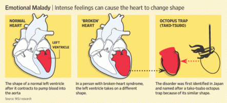

Sex after a heart attack: why many avoid it
By Deborah Kotz : Boston Globe : July 29, 2013
After going through the experience of a life-threatening heart attack, many patients are justifiably terrified of having another — perhaps one they won’t survive — and some avoid sex for this reason. If their doctors took the time to discuss the resumption of sexual activity, however, women recovering from heart attacks have reported in a new study that they’d be far more likely to return to their former sex lives.
Unfortunately, very few cardiologists broach the topic of sex, especially with their female patients, according to research published last week in the Journal of the American Heart Association. Researchers extensively interviewed 17 female heart attack patients and found that very few of their doctors discussed resuming sex unless the women asked directly for guidance.
“These patients told us it would be easier to overcome their fears of sex after having a heart attack if their doctors gave them more information,” said study co-author Stacy Lindau, a gynecologist at the University of Chicago Medicine who specializes in treating sexual dysfunction in people undergoing cancer and other medical treatments.
“Even providing just one sentence of counseling on this issue is so much more than saying nothing and can have a real impact on patient outcomes,” she added.
The new study builds on previous research also conducted by Lindau involving 1,900 heart attack patients who were surveyed about their sex lives after leaving the hospital. That study published last year found that only one-third of women and slightly less than half of men received hospital discharge instructions about resuming sexual activity. Those who didn’t get any advice were 44 percent more likely to report a year later that they still hadn’t resumed intercourse.
The American Heart Association issued landmark recommendations in 2012 advising cardiologists to discuss the resumption of sexual activity with heart attack patients. In the majority of cases, the panel of experts concluded, a heart patient’s risk of having a heart attack during sex was no greater than the risk faced by their peers of the same age without any heart problems.
A number of factors could explain why physicians and nurses fail to follow these recommendations, said Elaine Steinke, a professor of nursing at Wichita State University in Kansas who helped write the 2012 recommendations. “Medical providers may feel embarrassed or not view it as important enough information to include in a conversation before a patient is discharged from the hospital.”
But, she added, doctors can easily fold it into any recommendations they provide on physical activity. The Heart Association guidelines said the stress to the heart during sex is equivalent to climbing two flights of stairs.
New recommendations concerning the specific type of sex counseling doctors should provide are set to be released by the association on Monday.
In the new study, some participants mentioned that their sexual activity was less frequent but that their physical intimacy with their partner — all were in committed relationships — had increased since their heart attack.
Others said having sex after their heart attack was life-affirming and helped them regain a sense of normalcy.
But many also had more fear and anxiety about how their bodies would handle the exertion, getting worried if their heart starting racing during foreplay. Spouses worried too. One woman said she had to convince her husband that she “wouldn’t die in bed.”
Another said she didn’t resume sex because she lost her libido, a side effect she attributed to the antidepressants she started taking to manage depression following her heart attack.
“Any sort of invasive procedure on any part of the body has the potential to affect someone’s sexual function,” Lindau said. “If a healing wound is painful, patients will be more protective of that part of the body and avoid putting pressure on it, which may require new positions for sexual intercourse.”
While cardiologists may not want to delve into this much detail with their heart attack patients, they should make referrals to health care providers who do, Lindau emphasized.
“We’ve seen from this study and previous ones that patients — especially women — who aren’t counseled about sex after a heart attack are less likely to resume it,” Steinke said. “These findings are a call for health care providers to do more. They must make this part of a routine practice.”
